EBV was the first human virus that was associated with cancer when in 1964 it was isolated from the cells of the tissue line of Burkitt's lymphoma.
It ranks among the nine human herpesviruses and as well as the others, has a lethal and productive (lytic) phase of the cell cycle, latent in B - lymphocytes and productive in epithelial cells. It is transmitted by saliva and in some cases was detected also in genital secretions of both genders which led to speculation about a possible sexual transmition.
Primary infection is most common in childhood, very often subclinical, or later during adolescence. 50 % - 74 % of cases are with a clinical image of mononucleosis considering the typical way of transmition called the "kissing disease". Seroprevalence in adults is more than 90 % with a long-term latent persistence of the virus in B-lymphocytes of which in approximately 20 % of individuals EBV is located in saliva. Another possible way of transmition of the viruses is the transfusion or transplantation due to its latent presence in B-lymohocytes of healthy donors.
Nowadays, it is associated with a variety of benign and malignant diseases of lyphatic tissues and epithelium. The incidence of some of them is bound to a particular ethnic group or geographic area. This includes for example infectious mononucleosis, Burkitt's lymphoma, Hodgkin's lymphoma, some other non-Hodgkin's lymphoma, lymphoproliferative disorder of boys with immunodeficiency linked to the X chromosome. From other diseases it is nasopharyngeal carcinoma etc..
EBV plays a role also in the formation of lymphoproliferation in other groups of patients with a compromised immune system such as iatrogenic immunosupressed people after a transplantation in whom may occur the posttransplantation lymphoproliferative disease – PTLD, in patients with HIV or in some patients with autoimmune diseases receiving immunosuppressive therapy.
In HIV positive patients EBV infection is besides that, the cause of the "hairy" leukoplakia in the oral cavity.
Interdependencies of the presence of EBV, grades of the disease and changes of immunity in post-transplantation lymphoproliferation, captured in figure 1.
-
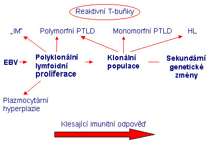 Fig.1
Fig.1Influence of EBV
Ebstein-Barr virus belongs to the gamma subfamily of potentially oncogenic herpesviruses which are distinguished by two groups - gamma1, Lymphocryptovirus, (LCV), and gamma2, Rhadinovirus (RDV).
EBV is human LCV and due to the differences in the sequences of some genes it distinguishes EBV-1 and EBV-2 with different geographical occurence but, in functional terms, without a significant difference.
The architecture of virion is similar to other herpesviruses. The annular core-protein coated by the DNA in nucleocapsid consisting of 162 capsomers is protected by a protein shell and outer shell with surface glycoprotein molecules.
EBV genome is composed of the linear double-stranded DNA of the size of 184 kb with 60 % content of GC pairs. According to the degree of latent or lytic infection it leads to transcription and translation of corresponding genes for some of the EB nuclear antigens EBNA-1, -2, -3A, -3B, -3C, -LP, for regulatory proteins ex. BZLF, BRLF1, integral membrane proteins LMP1, -2A, -2B, for the capside and envelope proteins and also the transcription of the genes for EBER-1,-2 (small nonpolyadenylated sequence of RNA) which are then translated only partially.
Expression of EBV genes is reduced in various degrees of latency when viral proteins serve to maintain episome in the cell, inhibit apoptosis and processing of antigens.
Examination
In our laboratory we use the following detection systems:
- Detection of the gene for nuclear antigen EBNA-1. The protein expressed from this gene can be detected in all cases of EBV-associated diseases. We use nested PCR which from the target region amplifies a product of the size of 297 bp using outer primers and in the second step inner primers amplify a product of the size of 209 bp.
- Detection of the gene for nuclear antigen EBNA-3C. One-step PCR amplifies the segment of the size of 153 bp.
- EBER in situ hybridization: EBV in situ hybridization is run on the BenchMark automated slide staining system (Ventana Medical System) using the INFORM EBER fluorescein-labeled oligonucleotide probe and iViewBlue detection kit (Ventana, Roche, Basel, Switzerland ) in order to detect the early RNA transcript of Epstein-Barr virus..
Analytical sensitivity: 10000 copies/ml.
References
- Herbst H, Dallenbach F, Hummel M, Niedobitek G, Pileri S, Muller-Lantzsch N, Stein H. Epstein-Barr virus latent membrane protein expression in Hodgkin and Reed-Sternberg cells. Proc Natl Acad Sci USA. 1991;88(11):4766-70.
- Anagnostopoulos I, Hummel M, Finn T, Tiemann M, Korbjuhn P, Dimmler C, Gatter K, Dallenbach F, Parwaresch MR, Stein H. Heterogeneous Epstein-Barr virus infection patterns in peripheral T-cell lymphoma of angioimmunoblastic lymphadenopathy type. Blood. 1992;80(7):1804-12.
- Anagnostopoulos I, Hummel M, Kaudewitz P, Korbjuhn P, Leoncini L, Stein H. Low incidence of Epstein-Barr virus presence in primary cutaneous T-cell lymphoproliferations. Br J Dermatol. 1996;134(2):276-81. Fields. Virology (fourth edition). Lippincot Williams & Wilkins, 2001.
- Hopwood P, Crawford DH. The role of EBV in post-transplant malignancies: a review. J Clin Pathol. 2000;53(4):248-54. Review.
- Fields. Virology (fourth edition). Lippincot Williams & Wilkins, 2001
- Macsween KF, Crawford DH. Epstein-Barr virus-recent advances. Lancet Infect Dis. 2003;3(3):131-40. Review.