Product of the gene HER2/neu (erbB2) is the transmembrane receptor with tyrosine kinase activity with a molecular weight 185 kDa that belongs to the family of the genes HER - homologs of EGFR (epidermal growth factor receptor). This gene is located on the long arm of the chromosome 17 in the region q11.2 - q12. HER2/neu gene amplifiation is found in many cancers. In mammary gland cancer this amplification is detected in 15 – 25 % of cases and strongly correlates with the overexpression of the corresponding protein.
Overexpression of the protein HER2/neu is an important prognostic and predictive factor of the invasive carcinoma of the mammary gland. It was found that this overexpression is associated with a worse prognosis. However, in patients with a proven HER2/neu gene amplification it is possible to use (albeit still with a questionable benefit) the treatment with Herceptin, a monoclonal antibody against the extracellular domain of the protein HER-2/neu.
Examination
Determination of the HER2/neu gene apmlifcation can be performed by a variety of the methodologies. Currently, the most used is fluorescent in situ hybridization (FISH). In our laboratory we use diagnostic PathVysionTM HER2/neu DNE probe kit (Vysis/Abbott).
-
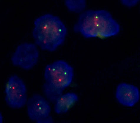 Fig.1a
The examination of HER2/neu amplification using FISH, performed on the Formalin-Fixed and Paraffin-Embedded (FFPE) tissues with directly labeled probes HER2/neu (red) and CEP17 (green). HER2/neu is amplified if the ratio between the number of HER2/neu and CEP17 is > 2.
A) without amplification -
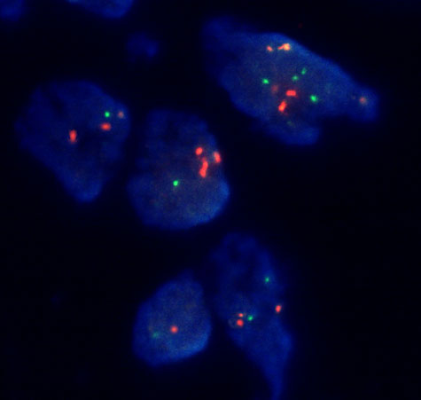 Fig.1b
The examination of HER2/neu amplification using FISH, performed on the Formalin-Fixed and Paraffin-Embedded (FFPE) tissues with directly labeled probes HER2/neu (red) and CEP17 (green). HER2/neu is amplified if the ratio between the number of HER2/neu and CEP17 is > 2.
B) slight amplification -
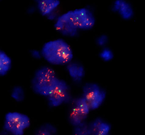 Fig.1c
The examination of HER2/neu amplification using FISH, performed on the Formalin-Fixed and Paraffin-Embedded (FFPE) tissues with directly labeled probes HER2/neu (red) and CEP17 (green). HER2/neu is amplified if the ratio between the number of HER2/neu and CEP17 is > 2.
C) extensive amplification
References
- Slamon DJ, Clark GM, Wong SG, Levin WJ, Ullrich A, McGuire WL. Human breast cancer: correlation of relapse and survival with amplification of the HER-2/neu oncogene. Science. 1987;235(4785):177-82.
- Kallioniemi OP, Kallioniemi A, Kurisu W, Thor A, Chen LC, Smith HS, Waldman FM, Pinkel D, Gray JW. ERBB2 amplification in breast cancer analyzed by fluorescence in situ hybridization. Proc Natl Acad Sci U S A. 1992;89(12):5321-5.
- Olayioye MA, Neve RM, Lane HA, Hynes NE. The ErbB signaling network: receptor heterodimerization in development and cancer. EMBO J. 2000;19(13):3159-67.
- Beyser K, Reiser A, Gross C, Moeller C, Tabiti K, Rüschoff J. Real-time Quantification of HER2/ neu Gene Amplification by LightCycler Polymerase Chain Reaction (PCR) - a New Research Tool Roche Molecular Biochemicals Biochemica 2001;2: 15-8.
- Slamon DJ, Leyland-Jones B, Shak S, Fuchs H, Paton V, Bajamonde A, Fleming T, Eiermann W, Wolter J, Pegram M, Baselga J, Norton L. Use of chemotherapy plus a monoclonal antibody against HER2 for metastatic breast cancer that overexpresses HER2. N Engl J Med. 2001;344(11):783-92.
- Wang S, Hossein Saboorian M, Frenkel EP, Haley BB, Siddiqui MT, Gokaslan S, Hynan L, Ashfaq R. Aneusomy 17 in breast cancer: its role in HER-2/neu protein expression and implication for clinical assessment of HER-2/neu status. Mod Pathol. 2002;15(2):137-45.
- Bernard PS, Wittwer CT. Real-time PCR technology for cancer diagnostics Clinical Chemistry 2002;48 (8): 1178-85.