Translocation t(14;18) (q32;q21) is one of the best-characterized cytogenetic abnormalities in B lymphoproliferative diseases. It is a typical marker of follicular lymphomas and to a lesser extent also in diffuse large B-cell lymphoma.
When chromosomal translocation t(14;18) (q32;q21), it comes to a fusion of the area surrounding one of the JH gene regions for the heavy chain of immunoglobuline (IgH) on the chromosome 14 and the bcl-2 gene on the chromosome 18. When this fusion occurs, the antiapoptotic gene bcl-2 gets under the influence of the enhancer for the IgH gene. As a consequence of this it comes to the deregulation of its normal expression and to the disruption of apoptotic processes. Breakages in the bcl-2 gene occurs approximately in 70 % cases of major breakpoint region (MBR) localized in 3' untranslated region of the exon 3 of this gene and then in the region called minor cluster region (MCR) localized about 20 kb distally from MBR.
Examination
Translocation t(14;18) (q32;q21), like many other translocations, is relatively easy to detect by molecular biological and cytogenetic methods. Currently, the most used methods are apparently that which are based on the various versions of PCR (polymerase chain reaction) and FISH (fluorescence in-situ hybridisation).
In our laboratory, we use one-round PCR with the set of primers overlapping MBR and MCR. In the case of positive sample, primers amplify a fragment of a variable length detectable by the agarose gel electrophoresis (fig. 1)
FISH
For the interphase FISH we use commercially available translocation probe IgH/BCL2 Dual Color, Dual Fusion Translocation Probe from the Vysis/Abbot company. With this probe we get two red and two green fluorescent signals in the normal nucleus (fig. 2A). In the sample with positive translocation t(14;18) (q32;q21) there are two yellow fused signals from translocated chromosomes and one red and one green signal from intact chromosomes (fig. 2B).
-
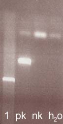 Fig.1
Fig.1Agarose gel with the PCR fusion products detecting translocation t(14;18) (q32;q21). MBR multiplex.
-
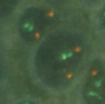 Fig.2A
A) Nucleus negative for translocation t(14;18) (q32;q21)
-
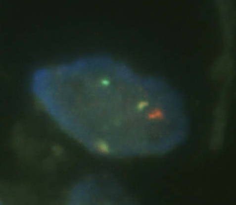 Fig.2B
B) Nucleus positive for translocation t(14;18) (q32;q21)
References
- World Health Organisation Classification of Tumours: Pathology and Genetics. Tumours of Haematopoietic and Lymphoid Tissues. Ed. Jaffe, ES, Harris, NL, Stein, H, Vardiman JW. IARC Press, Lyon, 2001.
- van Dongen JJ, Langerak AW, Bruggemann M, Evans PA, Hummel M, Lavender FL, Delabesse E, Davi F, Schuuring E, Garcia-Sanz R, van Krieken JH, Droese J, Gonzalez D, Bastard C, White HE, Spaargaren M, Gonzalez M, Parreira A, Smith JL, Morgan GJ, Kneba M, Macintyre EA. Design and standardization of PCR primers and protocols for detection of clonal immunoglobulin and T-cell receptor gene recombinations in suspect lymphoproliferations: report of the BIOMED-2 Concerted Action BMH4-CT98-3936. Leukemia. 2003;17(12):2257-2317.